Pacemaker Implantation

What is a Pacemaker?


Pacemaker is a small device implanted under the skin to regulate heart rhythm by sending electrical pulses to the heart muscle, ensuring it beats regularly and efficiently. It’s used to treat arrhythmias and improve overall heart function.
 
The main function of a pacemaker is to monitor the heart’s electrical activity and intervene when it detects irregularities. The heart relies on electrical impulses to contract and pump blood effectively. In some individuals, these impulses may become irregular or too slow, leading to symptoms such as dizziness, fainting, shortness of breath, or fatigue. A pacemaker helps by sending electrical impulses to the heart muscle, ensuring it beats at a regular rhythm and maintains an adequate heart rate.
 

Components of a Pacemaker:

Pulse Generator: This is the main component of the pacemaker and contains the battery and electronic circuitry. The battery supplies power to the pacemaker and can last several years depending on usage.
 
Leads: These are thin, insulated wires that carry electrical impulses from the pulse generator to the heart and transmit information about the heart’s activity back to the pulse generator. Modern pacemakers can have one to three leads depending on the type of arrhythmia being treated.
 
Sensing Electrodes: These are part of the leads and detect the heart’s natural electrical signals. The pacemaker uses this information to determine when and how to deliver electrical impulses.
 
Programmer: This is an external device used by healthcare providers to program and adjust the settings of the pacemaker. It allows customization of the pacing parameters based on the patient’s specific needs.


How a Pacemaker Works:

 
Sensing: The pacemaker continuously monitors the heart’s electrical activity through the sensing electrodes. It detects the natural heartbeat and determines if additional pacing is needed.
 
Pacing: If the pacemaker detects a slow heart rate or an abnormal rhythm, it delivers a small electrical impulse to the heart muscle through the leads. This causes the heart to contract and maintain a regular rhythm.
 
Rate-Responsive: Some pacemakers are designed to adjust the heart rate based on the body’s needs. They can increase heart rate during physical activity and decrease it during rest.
 
Monitoring: Pacemakers can store information about the heart’s activity, which can be retrieved during follow-up appointments to assess how well the device is functioning and to make any necessary adjustments.


Types of Pacemakers:

 
Single-Chamber Pacemakers: These have one lead connected either to the right atrium (upper chamber) or the right ventricle (lower chamber) of the heart.
 
Dual-Chamber Pacemakers: These have two leads—one in the right atrium and one in the right ventricle. They coordinate the timing of electrical impulses between the two chambers to mimic the heart’s natural rhythm more closely.
 
Biventricular Pacemakers (Cardiac Resynchronization Therapy, CRT): These are used in patients with heart failure and have three leads—one in the right atrium and two in the right and left ventricles. They help synchronize the contractions of the heart’s lower chambers to improve pumping efficiency.

The Benefits of Pacemaker Implantation

Pacemaker implantation offers significant benefits for individuals with certain cardiac conditions characterized by abnormal heart rhythms or slow heart rates (bradycardia). Here are the key benefits:
 
Restoration of Normal Heart Rate: Pacemakers ensure that the heart beats at a regular rate, which is crucial for maintaining adequate blood flow to the body’s organs and tissues. By delivering electrical impulses to the heart muscle when needed, pacemakers prevent symptoms associated with slow heart rates, such as dizziness, fainting, fatigue, and shortness of breath.
 
Improved Quality of Life: For many patients, pacemaker implantation leads to a dramatic improvement in symptoms and overall quality of life. It allows individuals to resume normal daily activities without the limitations imposed by symptoms of bradycardia or irregular heart rhythms.
 
Symptom Relief: Pacemakers effectively alleviate symptoms caused by bradycardia, such as lightheadedness, weakness, and episodes of passing out (syncope). By maintaining a steady heart rate, these devices prevent interruptions in blood flow that can cause these distressing symptoms.
 
Enhanced Exercise Tolerance: Individuals with pacemakers often experience improved exercise tolerance and endurance. By ensuring the heart responds appropriately to physical activity, pacemakers enable patients to exercise regularly without experiencing symptoms of inadequate blood circulation or heart rhythm disturbances.
 
Prevention of Complications: Pacemakers help prevent potentially serious complications associated with untreated bradycardia or certain arrhythmias. These may include heart failure, stroke, or other cardiovascular events that can arise when the heart’s rhythm is irregular or too slow to meet the body’s demands.
 
Customizable Therapy: Modern pacemakers are programmable devices that allow healthcare providers to customize pacing parameters based on individual patient needs. This flexibility ensures optimal therapy tailored to each patient’s specific condition and physiological requirements.
 
Long-Term Management: Pacemakers are durable devices with batteries that typically last several years. Regular follow-up appointments are necessary to monitor the device’s function, adjust settings as needed, and replace the device when the battery eventually runs out. Long-term management ensures ongoing effectiveness in managing cardiac rhythm disorders.
 
Technological Advancements: Advances in pacemaker technology continue to improve device performance, longevity, and patient outcomes. New features such as rate responsiveness (adjusting heart rate based on activity level) and remote monitoring capabilities enhance patient safety and convenience.
 
Safety and Reliability: Pacemakers have a proven safety record and are considered a reliable treatment option for individuals with specific cardiac conditions. The procedure for implantation is generally safe, with low rates of complications when performed by experienced healthcare providers in appropriate clinical settings.

What to Expect After Pacemaker Surgery

After pacemaker surgery, patients can expect a period of recovery and adjustment as they adapt to the presence of the device and its impact on their daily lives. Here’s a comprehensive overview of what to expect:
 
Immediately After Surgery:
 
Hospital Stay: Most patients remain hospitalized for monitoring and recovery for 1-2 days after pacemaker implantation. During this time, vital signs are closely monitored, and any post-operative pain or discomfort is managed with medications.
 
Incision Care: Patients will have a small incision where the pacemaker was implanted, usually near the collarbone. It’s important to keep this area clean and dry to prevent infection. The healthcare team will provide instructions on how to care for the incision and when to remove any dressings.
 
Activity Restrictions: Initially, patients are advised to limit movement of the arm on the side where the pacemaker was implanted to allow the incision site to heal properly. Heavy lifting and strenuous activities should be avoided for a few weeks to prevent strain on the incision.


Recovery Tips for Pacemaker Patients

Recovery after receiving a pacemaker involves several key steps to ensure optimal healing and adjustment to the device. Here are some essential recovery tips for pacemaker patients:
 
Post-Procedure Care: Immediately after implantation, it’s crucial to follow the doctor’s instructions regarding wound care and activity restrictions. Keeping the incision site clean and dry helps prevent infection.
 
Activity Level: Initially, avoid strenuous activities and lifting heavy objects to allow the pacemaker leads to settle properly. Gradually resume normal activities as advised by your healthcare provider.
 
Medication Adherence: Take all prescribed medications regularly, especially those related to preventing infection or managing heart conditions.
 
Monitoring: Attend follow-up appointments to ensure the pacemaker is functioning correctly and to adjust settings if needed. Remote monitoring systems can also be set up for regular check-ups.
 
Driving Restrictions: Discuss with your doctor when you can safely resume driving, as regulations vary depending on location and individual circumstances.
 
Lifestyle Adjustments: Adopt a heart-healthy lifestyle, including a balanced diet low in sodium and saturated fats, regular exercise within recommended limits, and avoiding smoking and excessive alcohol consumption.
 
Emotional Support: Adjusting to life with a pacemaker can be challenging emotionally. Seek support from family, friends, or support groups to share experiences and address concerns.
 
Device Identification Card: Carry a pacemaker identification card at all times, which provides crucial information in emergencies and during security screenings.
 
Electromagnetic Interference: Be mindful of devices or environments that could interfere with the pacemaker’s function, such as strong magnets or certain medical procedures. Your healthcare provider can provide specific guidelines.
 
Alert Healthcare Providers: Inform all healthcare providers about your pacemaker before any procedures, tests, or treatments to avoid potential complications.
 
Watch for Signs of Complications: Be aware of symptoms like dizziness, chest pain, palpitations, or swelling around the device site, which could indicate issues needing immediate medical attention.
 
Education and Support: Educate yourself about your pacemaker model, its functions, and potential signs of malfunction. Knowledge empowers you to manage your health effectively.
 

Conclusion :

Choosing the best hospital for Pacemaker Implantation in Madinaguda involves considering factors such as the hospital’s reputation, the expertise of cardiologists and cardiac surgeons, the availability of advanced technology for diagnostics and procedures, patient outcomes, and the quality of post-operative care. Researching and consulting with healthcare professionals is essential to ensure you receive the highest standard of cardiac care tailored to your specific needs and preferences.

Ear Nose and Throat (ENT) Problems and Treatments

ENT Issues When to Consult an ENT Expert

We have to balance work and home in today’s fast-paced society. Therefore, it is even more crucial that we take additional care of our health. But frequently, especially when it comes to ENT (ear, nose, and throat) problems, we neglect to take the simple steps that our bodies require to remain healthy. Initially, we frequently have a tendency to ignore them or attempt home cures, which can occasionally make the situation worse and become a chronic illness.

These are some typical ENT problems that you should not put off seeking immediate attention from an ENT specialist.

Prolonged Sore Throat:

Mild illnesses or certain other conditions can cause sore throats. A few drugs or treatments may sometimes help them get better, but if the issue continues, it may raise some red flags. You should consult our ENT specialist in Madinaguda if you have been suffering from a painful throat for an extended period. Early treatment can prevent numerous consequences from developing from a sore throat, which can indicate many ailments and diseases.

Tonsil Issues :


An ENT expert should always be consulted for tonsil issues. Waiting for your home cures to take effect won’t help; in fact, many symptoms will only worsen with time. They could lead to issues like severe sore throats or trouble swallowing. It’s critical to visit a doctor since tonsil issues require prompt treatment.

Unexpected Deafness:

You might believe that your throat has nothing to do with your unexpected loss of hearing. In actuality, though, your throat, nose, and ears are all connected. Hearing loss requires emergency medical attention since it may indicate more serious underlying issues. Visit our Hospital for ENT treatment from one of the top ENT Doctors in Madinaguda if you have abrupt hearing loss or other hearing problems.

A Lump in the Neck :

A lump in the neck may indicate more serious medical conditions including thyroid or glandular problems. It may potentially be an indication of malignancy. Therefore, you need to consult a physician as soon as you discover a lump in your neck. You can recover and prevent serious consequences with the correct care and drugs.

Sinus Ache:

An ENT specialist is necessary for the treatment of sinus pain, which might indicate many different diseases. Common colds, viral infections, sore throats, and a host of other issues are associated with sinuses. The best course of action is to have this investigated because the appropriate care can improve your quality of life and make it more comfortable for you.

The Hospital for ENT Treatment at Archana Hospital, Madinaguda offers state-of-the-art equipment, state-of-the-art diagnostic facilities, and some of the best ENT doctors in the area to provide the best care. You can get relief from serious ENT difficulties such as sinus problems, ear pain, throat problems, and more with our cutting-edge diagnostic and surgical technology. To learn more about our offerings and facilities, please get in contact.

Valve Replacement Surgery: Types, Procedures, And Recovery

Valve Replacement Surgery: Types, Procedures, And Recovery

A form of heart illness called valvular Heart Disease arises when one or more of the four heart valves malfunction. Surgery to replace the heart valves may be necessary if they are too brittle, damaged, or otherwise unfit to mend. This procedure can be necessary if you have an aortic valve problem.
 
Blood flows from your heart into your aorta, which is your body’s main artery when this valve opens. When it shuts, blood flows to the rest of your body. When your aortic valve closes, it stops blood from flowing backward into your heart. It repeats this cycle with every pulse. Your physician might suggest replacing the valve surgically.

Why Could I Require Surgery To Replace Or Repair A Heart Valve?

Surgery to replace or repair one or more damaged heart valves is done to address the problems they cause.
 
The following symptoms could indicate heart valve injury or disease:
 

What Are The Risks Of Having Surgery To Replace Or Repair A Heart Valve?

The following hazards could arise with heart valve replacement or repair surgery:
 
> Bleeding during or after the surgery
> Blood clots that can cause lung problems, heart attacks, or strokes
> Virus Infection
> A case of pneumonia
> Pancreatitis
> Breathing difficulties

Types Of Heart Valve Surgery

The following are the two kinds of heart valve surgery:
 
> A large portion of the patient’s tissue is preserved during valve repair surgery, which replaces the damaged or faulty valve. The mitral valve is repaired most frequently, while problems with the aortic and tricuspid valves can also be addressed with repair surgery.
 
> Surgery to replace the damaged valve with a biological (derived from pig, cow, or human tissue) or mechanical (made of metal or carbon) valve. Your body won’t reject the replacement valve because every valve replacement is biocompatible.
 
To identify the kind, location, and degree of your valve ailment, your healthcare provider will test you. The test results are used to choose the best procedure for you. The following will also be considered by your physician:
 
 
Your cardiac surgeon may choose to combine valve surgery with other cardiac procedures. Examples include combining valve surgery with: and performing several valve surgeries simultaneously during surgery.
 
> Avoid surgery
> Procedure to treat an aortic aneurysm
> Surgical treatment for atrial fibrillation
Medicines And Food
Speak with your healthcare provider about:
 
> When and how should you take your regular medications before surgery?
> When should you stop eating and drinking before surgery?
 
Personal Goods And Clothing
 
Your healthcare providers may suggest that you bring several items to the hospital, including:
 
What Happens After A Heart Valve Operation?
 
After surgery, your medical team may move you to an intensive care unit (ICU) so they can keep a close eye on you. After that, you’ll be in a typical room. Five to seven days could pass while you’re in the hospital.
 
There will be gadgets linked to you to monitor your heart rate and blood pressure. You may also have tubes going out of your chest to drain fluid.
 
Your surgeon will advise you to resume eating, drinking, and exercising as soon as possible following surgery. You might start with short walks in your room or down the hallway and work your way up to a larger distance.
 
Your physician could advise you to sign up for cardiac rehab, which is a strictly monitored workout regimen.
Recovery
 
Complete healing could take many weeks or even months, depending on your healing pace and the type of operation you had. Keeping your incisions clean is essential because infection is the major concern right after surgery. Never delay in contacting your physician if you see any of the following symptoms of a disease:
 
fever, chills, soreness or edema near the wound, and a buildup of wound drainage.
 
When ought I to see my physician?
 
Consult your physician if
 
> Your chest or the region surrounding your incision pain.
> You go through depressive episodes. This can happen after surgery and make recovery take longer.
> You may have an infection if you have a fever.
> Your weight has increased by more than five pounds, which suggests fluid retention.

Best cystoscope Hospital in Madinaguda – cystoscope

What is cystoscopy?


A cystoscopy is a process used by medical professionals to see inside the bladder and urethra (the tube that removes waste from the body). It is carried out by a urologist using a lit instrument called a cystoscope that has a camera or viewing lens.

What are the types of cystoscopes?

Two varieties of cystoscopes exist:

Flexible cystoscope: Your doctor may inspect the interior of your bladder and urethra by carefully following the natural course of your urethra with a flexible scope.

Rigid cystoscopes: They don’t bend. Instruments for biopsies or tumor removal may be passed via the tube by your healthcare professional. Usually, this cystoscope is only utilized under general anesthesia or sedation.

The one that functions best for your particular treatment will be used by your healthcare professional.

When should I know the results of cystoscopy?

Your provider might not get the results of your biopsy for up to two weeks. Your practitioner will be aware of the outcome as they proceed if they are solely performing a cystoscopy and no other treatments concurrently. Find out from your provider when you can expect to receive a notification or see your results in your records.

When should I call my healthcare provider?

The majority of post-procedure issues, such as painful urination and bloody urine, resolve in a day or two. If issues persist longer than expected or if you encounter:

> Intense pain with urination.

> Significant blood flow or clots in your urine.

> A painfully full bladder and difficulty urinating.

> Symptoms of infection (pelvic discomfort, fever, foul-smelling or hazy urine).

Preparation Before Cystoscopy

Before performing a cystoscopy, a urologist may question a patient about any allergies, past medical history, and pre-procedural measures. The physician might advise:

> Before the surgery, stop using medications, such as blood thinners.

> When to eat and drink

> When to empty the bladder before the procedure or to give a urine sample for laboratory test

Who needs a Cystoscopy?

A cystoscopy may be advised by the physician if a patient encounters:

> Problems passing pee or controlling urination

> Stone in the bladder

> Blood in the pee or hemoptysis

> Dysuria, or pain during urinating

> Urinary tract infection that keeps coming back

What are the potential risks or complications of a cystoscopy?

Cystoscopy procedures are generally low-risk. Possible issues consist of:

> Infection (UTI).

> Contractions of the bladder. These may result in leaking urine and excruciating pains.

> Harm to your bladder.

> Harm to your urethra, such as constriction or scarring.

Side Effects of Cystoscopy

Following a cystoscopy, some adverse effects include:

> Urine with blood in it, usually just a few drops

> Burning in the urine, discomfort, and frequent urination for a few hours

Side effects peak to fade after 48 hours. See a physician if the issues persist longer and result in really painful urinating.

Conclusion:

Archana Hospitals in Madinaguda stands out as the premier destination for cystoscopy procedures. Renowned for its advanced facilities and skilled medical professionals, it ensures top-notch care and accurate diagnostics. Patients receive comprehensive treatment, coupled with compassionate support, ensuring optimal outcomes and peace of mind. Trust Archana Hospitals for unparalleled excellence in cystoscopy procedures.

ACL Repair Causes, Treatment, Repair & Recovery

ACL Repair Causes, Treatment,

 Repair & Recovery

One important ligament that stabilizes the knee joint is the Anterior Cruciate Ligament (ACL). Its function is to stop the tibia bone from moving forward too much about the femur bone. One common sports injury that needs surgical repair is an ACL tear. This blog will discuss the causes, care, and recovery of an ACL reconstruction.

 Causes of ACL Tears :

ACL tears can happen for several reasons, including:

Sports injuries: Because high-impact sports like football, basketball, and soccer require quick stops, pivots, or direction changes, players are likelier to suffer an ACL tear.

Trauma: Other traumatic occurrences that put undue strain or tension on the knee joint, such as falls, auto accidents, or other collisions, can also result in an ACL tear.

Overuse: Activities like long-distance running and jumping that repeatedly put stress on the knee joint raise the possibility of an ACL tear.

Treatment Options for ACL Tear

The degree of the injury determines how an ACL tear is treated. While non-surgical therapy options could be enough in some circumstances, surgery might be necessary in others.

1. Non-surgical treatment: Physiotherapy, pain management, and rest, ice, compression, and elevation (RICE) therapy are examples of non-surgical treatment alternatives. Protocol-Oriented Regaining functional independence requires physiotherapy, and patients with ACL injuries are always encouraged to perform these exercises to strengthen their knees.

2. Surgical intervention: Surgery is required when there is a high-grade ACL injury and noticeable indications of knee instability. During ACL repair surgery, a tissue graft—typically derived from the patient’s patellar tendon or hamstring—is used to reconstruct the injured ligament. The process is carried out as keyhole Surgery

Recovery from ACL Reconstruction Surgery

Professional athletes recuperate from ACL reconstruction surgery differently than non-professional players. It is advised that professional athletes wait six months before returning to competition, but this can be shortened to four months.  

Patients can usually get back to their regular activities in 3–5 weeks, and they can return to sports in 4-6 months. Physical therapy is required to restore the knee joint’s strength, flexibility, and range of motion throughout the healing phase.

In conclusion, a typical sports injury that can be quite crippling to the targeted athlete is an ACL tear. Depending on how severe the injury is, there are non-surgical and surgical treatment options. The recovery period following ACL reconstruction surgery can range from six weeks to three months. Patients who receive precise surgical care, appropriate post-operative care, and well-coordinated physiotherapy can restore complete functioning to their knee joint. 

Archana Hospitals excels in ACL reconstruction surgery, offering state-of-the-art facilities and expert surgeons. Patients receive personalized care, innovative techniques, and comprehensive rehabilitation, ensuring optimal recovery and restored knee function. Trust Archana Hospitals for exceptional outcomes in ACL reconstruction.

Hemorrhoidectomy

Hemorrhoidectomy


The surgical treatment used to eliminate hemorrhoids is called a hemorrhoidectomy. Hemorrhoids usually don’t require surgery, but if they persist or create significant problems, a hemorrhoidectomy can permanently remove them. The process is straightforward, although recuperation could be a little challenging. Take our recommendations to reduce discomfort and avoid constipation.

Overview

What is a Hemorrhoidectomy?

The surgical treatment used to eliminate Hemorrhoids is called a hemorrhoidectomy. Enlarged blood vessels in the anus (butthole) known as hemorrhoids can occasionally result in painful symptoms including bleeding and anal pain.

Hemorrhoids are quite frequent and often not harmful. Most won’t even need surgery or other medical care. However, a hemorrhoidectomy may be the best course of action for some hemorrhoids and in some situations.

Risks / Benefits

What benefits does hemorrhoidectomy offer in comparison to other hemorrhoid treatments?

The main benefit is that it functions. Surgically excised hemorrhoids usually don’t recur. Surgery should ultimately provide you with permanent relief, even though the recovery period is longer than with other treatments.

Another crucial option in an emergency is a hemorrhoidectomy. Surgery can be used to treat a thrombosed or strangulated hemorrhoid quickly and avoid worsening the condition.

What makes a Person get a Hemorrhoidectomy?

If your hemorrhoids are more complicated than usual, your doctor may suggest hemorrhoidectomy surgery. The following complications could necessitate a hemorrhoidectomy:

Prolapse: This indicates that your hemorrhoids have descended from inside your anus and are now protruding from the orifice, increasing the risk of irritation, bleeding, or injury. Prolapse is a progressive condition that often gets worse with time. Initially, Grade II hemorrhoids may only momentarily protrude from your anus during a bowel movement. However, your provider could advise removing them if you have to manually put them back in (Grade III) or if they won’t go back in at all (Grade IV).

Thrombosis: This is the point at which the hemorrhoid has swelled to the point where blood clots inside it, stopping the flow of blood. The color of thrombosed hemorrhoids is purple-blue. They may cause excruciating agony. They may occasionally burst, heavily bleeding. It will eventually heal on its own if you do not seek treatment straight away. But you might prefer surgery during the excruciating first 72 hours.

Constriction Incarcerated hemorrhoids are another name for Grade IV hemorrhoids that have prolapsed and refused to retract. A hemorrhoid in prison runs the risk of strangling itself. When something is strangled, it indicates that its circulation has been cut off and it is stuck in a constricted area. There is an urgent situation. In contrast to thrombosis, strangulation is not a self-limiting illness. Gangrene results from the strangulated tissues’ eventual death.

ReturnHemorrhoids can recur after less intrusive treatments or don’t go away. This may have a significant long-term effect on your life quality. Hemorrhoidectomy has a 95% success rate when other therapies are unsuccessful. You may decide to get a hemorrhoidectomy to permanently remove these hemorrhoids, even if you don’t have any other difficulties.

What can I anticipate from my hemorrhoidectomy recovery?

Following a hemorrhoidectomy, you could anticipate varying degrees of:

Bleeding: Following a hemorrhoidectomy, some bleeding is typical, particularly after a bowel movement (poop). There may be blood on your toilet paper or in your feces. Along with some clear or yellow discharge, you can also notice some in your underpants. To assist in absorbing the liquid, you can use gauze or a sanitary pad. This may go on for a month or longer. There shouldn’t be much blood. If you strain too hard to void, you run the risk of splitting the wound open, which could lead to more serious bleeding. To avoid this, constipation must be avoided. Occasionally, some patients have acute bleeding following surgery.

Swelling: Following a hemorrhoidectomy, swelling is typical. Anus swelling might have a feeling akin to hemorrhoids. It’s possible that you still think of them or that they’ve returned, but these are false impressions. Occasionally, a skin tag or lump may appear where the wound was made, which can also be misleading. Usually, these disappear after you’ve healed. Everyone’s healing process is different. Applying ice wrapped in a towel to the incision a few times a day for ten minutes at a time will help minimize swelling. Lying on your stomach with a pillow under your hips can also be beneficial. Try to sleep in this manner.

Pain: After a hemorrhoidectomy, some pain is to be expected, and most patients report rather high pain levels. However, you ought to be able to use medicine to control your pain. Typically, medical professionals provide a range of pain management techniques, such as topical and oral drugs as well as gentle wound care. Start taking your drugs as soon as you feel any discomfort following your procedure to avoid it altogether. Most report that, after two weeks, the pain has subsided. Consult your healthcare practitioner if your discomfort persists for more than two weeks, if you find it difficult to manage, or if it keeps you from urinating or passing gas.

Recovery after Haemorrhoid Removal

Following your Hemorrhoid removal, you may be advised to do the following self-care measures:

Medication: Take your prescriptions exactly as directed by your physician. This could involve laxatives, antibiotics, and painkillers.

Activities: Make sure you get enough sleep, but also make time to be active. Walking is a wise decision.

Diet: A diet that reduces the likelihood of constipation may be advised by your doctor.

Additional actions could be using ice packs, taking stool softeners, taking a sitz bath, and drinking plenty of water.

What are the possible risks or complications of hemorrhoidectomy surgery?

Among the short-term hazards of surgery are:

Thrombi.

Negative effects of the anesthetic.

Infection of the wound.

Excessive loss of blood.

Among the issues that could arise when you’re recovering are:

Possible long-term complications that can develop include:

Gradual recovery.

Severe pain even after taking medicine.

Urinary retention due to pain when peeing.

Constipation due to pain when pooping.

Narrowing of your anal canal from excessive scarring (anal stenosis).

Difficulty holding in your bowel movements due to muscle or nerve damage (fecal incontinence).

With careful technique, serious complications are rare.

Who is not a candidate for a hemorrhoidectomy?

If you have specific medical conditions that could raise your risk of major complications from the treatment, your healthcare professional might not propose hemorrhoidectomy for you. Among them are:

Immunosuppression.

Inflammatory bowel disease (IBD).

Bleeding disorders.

Portal hypertension.

Kidney Stones Treatment in Hyderabad | and Surgery Treatment Options

Kidney Stones : What are the

 Treatment Options?

You may have a few therapy choices if kidney stones, also known as urolithiasis, have been identified in you. These include ureteroscopy, percutaneous nephrolithotripsy (PCNL), extracorporeal shock wave lithotripsy (ESWL), and medicinal therapy.

An overview of the urinary tract’s anatomy

The urinary tract consists of

> Kidneys, two organs that remove excess water and waste from the blood

> Ureters, the two tubes that each kidney uses to send urine to the bladder

> Bladder, the organ that holds pee

> The urethra is a single channel that the body uses to expel urine from the bladder.

The kidney stone assessment

An evaluation that includes imaging is frequently the first step if your symptoms point to kidney stones. An intravenous pyelogram (IVP), a form of abdomen x-ray, was the accepted standard of therapy for a long time. This is no longer used in the majority of hospitals; instead, unenhanced helical CT scanning is used. Renal ultrasound can be used as a substitute in some circumstances, such as when a person has decreased renal function or an allergy to contrast dyes.

Blood tests, such as those for renal function (creatinine, BUN), will also be administered to you. Further blood tests may also be recommended by your physician. If an infection is suspected, a urine culture will be provided along with the results of the urinalysis.

Treatment with Medication for Kidney Stones

According to the majority of data, stones smaller than 10 mm in diameter have a good possibility of naturally exiting the urinary system. You can be recommended to undergo medical expulsive therapy (MET) with an alpha blocker drug such as tamsulosin. It’s critical to recognize that this is an off-label use of the medication. Intraoperative floppy iris syndrome is a rare complication that tamsulosin and can occasionally develop and complicate cataract surgery.

The use of MET is still debatable, and not all specialists agree that it is beneficial. Talk to a urologist or your doctor about your options.

lithotripsy using Extracorporeal Shock Waves

Every shock wave lithotripsy device sends shock waves through the kidney stone’s skin. The stone receives most, but not all, of the energy from the shock wave.

The best indicator of ESWL success is stone size. In general:

> Stones smaller than 10 mm can be successfully treated with ESWL; 

> For stones between 10 and 20 mm, extra considerations including stone placement and composition should be made. 

> Stones bigger than 20 mm are typically not successfully treated with ESWL.

Because the shards of a lower third kidney stone may not be completely removed from the kidney following fragmentation, these stones can also be harmful. These kidney fragments don’t exit the kidney as quickly as kidney fragments from the middle and upper thirds because of gravity.

The effectiveness of ESWL treatment is also influenced by obesity. The skin-to-stone distance (SSD) will be computed by the urologist to assess the likelihood of treatment success.

Among the potential side effects of ESWL

> Hemostases, or bruises to the kidney tissue, can happen occasionally but normally go away on their own with no further care.

> In the ureter, fragmented stones may build up and become obstructed. This is referred described as a “street of stones,” or steinstrasse. Any issues related to steinstrasse are frequently reduced by a ureteral stent. The procedure takes a few days or weeks to complete.

> Hypertension occurs in a tiny subset of patients receiving ESWL, while the exact mechanism is unknown.

There have also been reports of an elevated risk of diabetes mellitus after ESWL. Nevertheless, sizable population research conducted at the same institution did not support these findings.

Intraoperative nephrolithotripsy:

During percutaneous nephrolithotripsy, a surgeon makes a tiny incision in the lower back to access kidney stones under the supervision of ultrasound or fluoroscopic imaging. The stones are broken up by a power source, like a laser or ultrasonography, and then the pieces are removed from the kidney using an internal stent or external tube.

Greater kidney stones (2 cm or more), complicated stones, or lower pole renal stones greater than 1 cm are typically candidates for this treatment. Complications could include bleeding, infection, and harm to the organs nearby.

Ureteroscopy:

During a ureteroscopy, a surgeon inserts a tube into the ureter, possibly all the way up into the kidney, via the urethra and bladder. Using semirigid or flexible equipment, ureteroscopy provides the surgeon with a great view of the entire inside of the urethra. The stones are then broken apart under direct observation by the surgeon using a power source that is threaded up through the ureteroscope. The urologist may decide to insert a postoperative stent for a few days.

While they are rare, complications can include sepsis and ureteric damage or constriction.

Transforming Lives: Knee Replacement at Archana Hospitals, Chandnagar

Are you or someone you know struggling with knee pain that hinders daily activities? Knee replacement surgery could be the transformative solution you’ve been seeking. At Archana Hospitals in Chandnagar, we specialize in restoring mobility and enhancing the quality of life through advanced knee replacement procedures.

Understanding Knee Replacement

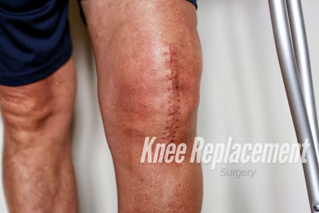
Knee replacement, also known as knee arthroplasty, is a surgical procedure aimed at replacing damaged knee joints with artificial ones. It’s typically recommended for individuals suffering from severe knee pain, stiffness, and decreased mobility due to conditions like osteoarthritis, rheumatoid arthritis, or injury.

Why Choose Archana Hospitals for Knee Replacement?

  1. Expertise: Archana Hospitals boasts a team of highly skilled orthopedic surgeons with years of experience in performing successful knee replacement surgeries. Our surgeons stay updated with the latest advancements in orthopedic care to ensure optimal outcomes for our patients.

  2. State-of-the-Art Facilities: Equipped with cutting-edge technology and modern infrastructure, Archana Hospitals provides a conducive environment for comprehensive knee replacement procedures. From pre-operative evaluations to post-operative care, we prioritize patient comfort and safety at every step.

  3. Personalized Care: We understand that every patient is unique, and their healthcare needs vary. That’s why we emphasize personalized care plans tailored to individual requirements. Our compassionate staff is dedicated to guiding patients through the entire treatment journey with empathy and support.

  4. Rehabilitation Services: Successful recovery from knee replacement surgery involves rehabilitation and physical therapy. At Archana Hospitals, we offer specialized rehabilitation programs designed to help patients regain strength, flexibility, and mobility post-surgery. Our multidisciplinary approach ensures holistic care for optimal recovery.

The Knee Replacement Process

  1. Initial Consultation: The journey begins with an initial consultation where our orthopedic specialists assess the patient’s medical history, symptoms, and diagnostic tests to determine the suitability for knee replacement surgery.

  2. Pre-operative Preparation: Before the surgery, patients undergo thorough pre-operative evaluations, including blood tests, imaging scans, and consultations with anesthesiologists. Our team educates patients about the procedure, addresses their concerns, and prepares them mentally and physically for the surgery.

  3. Surgical Procedure: The knee replacement surgery is performed under anesthesia, and the damaged portions of the knee joint are replaced with artificial implants made of metal, plastic, or ceramic. Our surgeons utilize minimally invasive techniques whenever possible to minimize surgical risks and accelerate recovery.

  4. Post-operative Care: Following surgery, patients are closely monitored in the recovery unit before being shifted to their rooms. Our medical team provides diligent post-operative care, including pain management, medication administration, wound care, and physiotherapy sessions to promote early mobilization and prevent complications.

  5. Rehabilitation and Follow-up: Rehabilitation plays a crucial role in optimizing the outcomes of knee replacement surgery. Our rehabilitation specialists work closely with patients to devise customized exercise regimens aimed at strengthening muscles, improving range of motion, and enhancing overall function. Regular follow-up appointments ensure that patients progress smoothly towards recovery milestones.

Experience Life Beyond Knee Pain

Don’t let knee pain limit your lifestyle and independence. Take the first step towards a pain-free future by exploring the advanced knee replacement services at Archana Hospitals, Chandnagar. With our unwavering commitment to excellence and patient-centric approach, we strive to empower individuals to reclaim their mobility and rediscover the joys of an active life. Contact us today to schedule a consultation and embark on your journey towards renewed vitality and well-being.

Cardiovascular Disease

Cardiovascular Disease

What is Cardiovascular Disease?

Cardiovascular Disease (CVD) refers to a range of conditions affecting the heart and blood vessels, including coronary artery disease, heart failure, and stroke. These ailments can manifest silently or with noticeable symptoms, posing serious health risks. At Archana Hospital, we’re committed to combating CVD with advanced treatments and expert care. Our team includes the Best Cardiology Surgeon in Madinaguda, offering unparalleled expertise in diagnosing and treating heart-related issues. Trust us for comprehensive cardiac care and a path toward better heart health. Your well-being is our priority.

Heart or blood vessel problems are included in cardiovascular disease, such as:

> constriction of blood arteries in your body 

>  heart, or in other organs.

> Issues with the heart and blood vessels exist from birth.

> Heart valves not functioning properly.

> Abnormal heartbeats.

Signs and Origins:

What is the root cause of heart disease?

Depending on the particular kind, there are many causes of cardiovascular disease. For instance, peripheral arterial disease and coronary artery disease are brought on by atherosclerosis, or plaque accumulation in the arteries. Arrhythmias can be brought on by drug side effects, hereditary disorders, coronary artery disease, or cardiac muscle scarring. Valve disorders can be brought on by rheumatoid arthritis, infections, and aging.

What are the risk factors for cardiovascular disease?

If you have any of the following risk factors, your chance of developing cardiovascular disease may increase:

> Elevated blood pressure, or hypertension.

> Hyperlipidemia, or high cholesterol.

> Usage of tobacco (including vaping).

> Diabetes type 2.

> Family history of heart disease.

> Lack of physical activity.

> Having excess weight or obesity.

> A Diet heavy in fat, sugar, and sodium.

> Excessive alcohol consumption.

> Abuse of illicit or prescribed drug use.

What Signs of Heart Disease are Present?

Symptoms of cardiovascular illness can differ according to the underlying reason. Older folks and people assigned female at birth may experience more modest symptoms. However, they can still have significant cardiovascular disease.

> Heart problems symptoms

Chest pain (angina).

> Pressure, heaviness, or discomfort in the chest that is > Occasionally referred to as a “weight on the chest” or a “belt around the chest.”

Breathing difficulty (dyspnea).

> Fainting or vertigo.

> Fatigue or fatigue.

Signs of Blood artery Blockages all Over your Body


> Walking causes pain or cramping in your legs.

> Leg ulcers that refuse to go away.

> Your legs’ skin may be cool or red.

> Enlargement of the legs.

> numbness in a leg or your face. You might just have this on one side of your body.

> Difficulty seeing, speaking, or moving.

How may Heart disease be avoided?

Certain forms of cardiovascular disease, such as congenital heart disease, are unavoidable. Lifestyle changes can, however, lower your chance of developing a variety of cardiovascular diseases.

You may lower your risk of cardiovascular disease by:

> Staying away from all tobacco products.

> Handling additional medical issues including High Blood Pressure, > High cholesterol, or Type 2 diabetes.

> Reaching a healthy weight and keeping it there.

> Eating a diet low in salt and saturated fat.

> Exercising for a minimum of 30 to 60 minutes most days.

> Lowering and controlling tension.

Note: 

Heart and blood vessel disorders are referred to as cardiovascular diseases. Heart attacks and strokes can result from heart disease if treatment is not received appropriately. Cardiovascular disease can be managed with medication or lifestyle modifications. An early diagnosis aids in the delivery of efficient care. Many people with cardiovascular disease have active, fulfilling lives.

Best Hospital in Hyderabad


Yashoda Hospital :

Yashoda Group of Hospitals has over the years emerged as a socially responsible corporate citizen. The Group is a silent crusader in the field of community services in its own dedicated way. The social initiatives of Yashoda Group are mostly centered on its core competency – Health. We are committed to the well-being of not just patients, but the entire community.
 

Kims Hospital :

Kims Hospital is one of the largest corporate healthcare groups in AP and Telangana in terms of the number of patients treated and treatments offered, according to the CRISIL Report. We provide multi-disciplinary integrated healthcare services, with a focus on primary secondary & tertiary care in Tier 2-3 cities and primary, secondary, tertiary, and quaternary healthcare in Tier 1 cities. We operate 9 multi-specialty hospitals under the “KIMS Hospitals” brand, with an aggregate bed capacity of 3,064, including over 2,500 operational beds as of December 31, 2020, which is 2.2 times more beds than the second-largest provider in AP and Telangana, according to the CRISIL report. We offer a comprehensive range of healthcare services across over 25 specialties and super specialties, including cardiac sciences, oncology, neurosciences, gastric sciences, orthopedics, organ transplantation, renal sciences, and mother & child care. 

Archana Hospitals :

ARCHANA HOSPITALS and its affiliates will be the healthcare provider of choice for physicians and patients – delivering the healthcare we expect for our own family – recognized for:

 
  • Exceptional quality
  • Unparalleled service and patient experience
  • Great place to work and practice
  • Academic programs and research that support core clinical missions
  • Clinically integrated affiliates

Best Cardiology Hospital in Hyderabad

Best Gynaecology Hospital in Chanda Nagar